Two Easy Ways to Start Earning Rewards!
Become a member today! Members-only pricing and offers, personalized care notifications, Vital Care points back on every purchase and more!
 Become a credit card member today!
Earn 2X Pals Rewards points at Petco
when you use Petco Pay!APPLY NOWLearn More About Petco Pay Benefits
Table of Contents
As a devoted pet parent, there have probably been times you’ve wished you could read your pet’s mind. Though you may be unable to ask your pet what’s wrong, you could have access to the next best thing—a dog or cat X-ray or ultrasound.
Veterinarians use diagnostic imaging like X-rays and ultrasounds to look inside a pet’s body. These diagnostic procedures are noninvasive and not painful. X-rays and ultrasounds can be used individually for certain health conditions or can even be used together to provide your veterinarian with even more information. What’s the difference between a cat or dog X-ray and a cat or dog ultrasound, and in what situations might either or both of these diagnostic tools be used? Keep reading to find the answers to some of your most pressing questions.
Ultrasounds and X-rays are just some of the many veterinary services offered by Vetco Total Care veterinarians.
What is an X-ray?
You probably know what a completed dog or cat X-ray image looks like—a black-and-white depiction of your pet’s skeletal structure and internal organs. But how does this detailed look at the inside of your pet get captured? X-ray images, also known as radiographs, are formed when electromagnetic waves of a particular frequency are passed through a part of your pet’s body. Different physical structures absorb X-ray waves at different levels, which is why various aspects of your pet’s anatomy appear clearly.
For instance, bones and metal absorb a lot of X-rays and therefore appear white on an X-ray image. Muscle and fat show up as varying shades of gray. Air appears black on the image because most X-rays pass through it. The finished cat or dog X-ray image is the result of the rays passing through the various tissues and structures of your pet’s body and forming a cohesive picture.
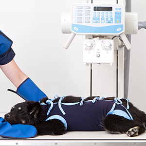
When are dog and cat X-rays used?
Veterinarians use X-rays as diagnostic aids. That means they’re not used to treat your pet’s health issue but to find out what might be causing it. X-rays aren’t used to help diagnose all health conditions but are invaluable in specific situations. They are frequently used to help diagnose problems affecting joints and bones, such as osteoarthritis, fractures, some spinal issues or infection and cancer.
If your pet has a limp, an X-ray can help determine whether conditions like a bone fracture or hip dysplasia might be the cause. Chest x-rays can help diagnose diseases that affect the heart, lungs and anything nearby. For example, an image of a dog or cats’ chest may indicate the presence of pneumonia, asthma or in some cases a tumor. Abdominal X-rays can identify problems in the gastrointestinal tract, urinary tract, liver, spleen and more. While certain dog vitamins and supplements or cat vitamins and supplements might be able to support your pet’s digestive health from the inside out, some more serious issues—such as intestinal blockages—may require X-ray imaging to diagnose.
Limitations
Though a cat or dog X-ray can be an invaluable diagnostic tool to help your veterinarian decide the best treatment for certain pet health problems, like all tools, the X-ray has its limitations. For instance, X-rays provide only a 2D snapshot of what’s happening in certain parts of a pet’s body— they are not dynamic video-style images that show your pet’s body functioning in real-time. That’s why they’re best used to diagnose conditions that can be properly displayed in a picture, such as a bone break, rather than, for instance, health issues that have to do with blood flow or heartbeat.
Another issue is that X-rays don’t do a great job differentiating between some internal structures. While larger cancerous masses could show up on a dog’s X-ray, a veterinarian probably wouldn’t be able to see a small tumor in an X-ray image of a dog’s liver because the tumor tissues and healthy liver tissues are too similar. Therefore, X-rays can’t be depended upon to diagnose all problems affecting your pet’s internal organs.
Finally, dog and cat X-rays require a pet to remain still while an image is taken. As a result, some pets might need to be sedated so that they stay still enough for your veterinarian to capture a quality image.
What is an ultrasound?
Like an X-ray, a dog or cat ultrasound is a diagnostic imaging tool that lets a veterinarian see inside your pet’s body. And like an X-ray, an ultrasound is created using waves—sound waves, in this case. Ultrasound images are made using a wand that passes sound waves above the frequency of normal human hearing. The ultrasound machine detects differences in the sound waves that are reflected back to the probe, and these differences are what create ultrasound images.
What’s more, your veterinarian can move the ultrasound wand to take images from different angles, giving a 3D look at your pet’s internal structures. Your vet can even take these images over time to monitor things like blood flow and heart movements, creating a more dynamic video-style image than is possible with a cat or dog X-ray.
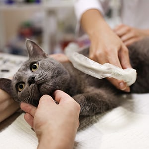
When are dog and cat ultrasounds used?
Cat and dog ultrasounds are frequently used to help diagnose problems that affect the heart, monitor blood flow through the heart and watch how the heart valves are working. This is called an echocardiogram—one of the most well-known ultrasounds a veterinarian might use on your pet. Meanwhile, if your vet gives your cat or dog an abdominal ultrasound, it’s so they can look at organs within your pet’s abdomen, such as the liver, kidneys, spleen, stomach, intestines and bladder. And of course, one of the most well-known uses of ultrasound technology is its use during pregnancy—which works in animals much the same as in humans. Ultrasounds can be used to identify whether a pet is pregnant, and while you might not be able to count how many kittens there are in a pregnant cat’s ultrasound, you can certainly confirm whether kittens are on the way.
Unlike X-rays, ultrasounds can differentiate between different types of soft tissue and can also tell the difference between soft tissue and fluid. For this reason, X-rays are more likely to be used in diagnosing bone breaks, while ultrasounds might be better suited for finding tissue tears. Ultrasounds can also be used to guide a biopsy needle to take tissue or fluid samples from inside the body.
How long does it take to do an ultrasound on a cat or dog?
If your pet needs an ultrasound, the procedure may be completed in about 30 to 60 minutes. However, the time it takes to complete a dog or cat ultrasound can depend on the body part being imaged, the condition being monitored and other factors. One thing most pet ultrasounds have in common is that unlike X-rays, they do not typically require a pet to be sedated during the imaging session, which means they can be completed relatively quickly and easily.
The limitations of dog and cat ultrasounds
A cat or dog ultrasound can be extremely useful to a veterinarian diagnosing a health condition that affects your pet’s soft tissues, but this helpful tool has its limitations. It may not be as useful as an X-ray for diagnosing certain problems, such as bone fractures and other skeletal issues.
The way an ultrasound is administered may also have some downsides. For instance, while ultrasound is generally known as a procedure that does not require pet anesthesia, some pets may need to be sedated to stay still during an imaging session. Additionally, a pet’s fur often needs to be shaved in preparation for a dog or cat ultrasound so that the wand can make contact with the skin. Ultrasound gel is also used in conjunction with the wand. Therefore, while ultrasounds aren’t invasive, they may require more direct interaction with your pet’s skin and fur than an X-ray would.